Nuclear medicine imaging uses small amounts of radioactive material, called a tracer, to create images of your organs and tissues. These pictures help diagnose and treat conditions like cancer, heart disease and other illnesses.
Advertisement
Cleveland Clinic is a non-profit academic medical center. Advertising on our site helps support our mission. We do not endorse non-Cleveland Clinic products or services. Policy
Nuclear medicine imaging is a test that uses small amounts of radioactive substances (called tracers) to diagnose or monitor diseases or illnesses. After you receive the tracer, a special camera senses the radiation it gives off as it moves through your body. Then, a computer produces detailed images of your organs and tissues.
Advertisement
Cleveland Clinic is a non-profit academic medical center. Advertising on our site helps support our mission. We do not endorse non-Cleveland Clinic products or services. Policy
Healthcare providers use these pictures to diagnose or treat many health conditions. Some of the conditions it can help diagnose are:
Nuclear medicine can treat certain conditions. It can also check how well a treatment is working. For example, it can help deliver radiation directly to a cancerous tumor.
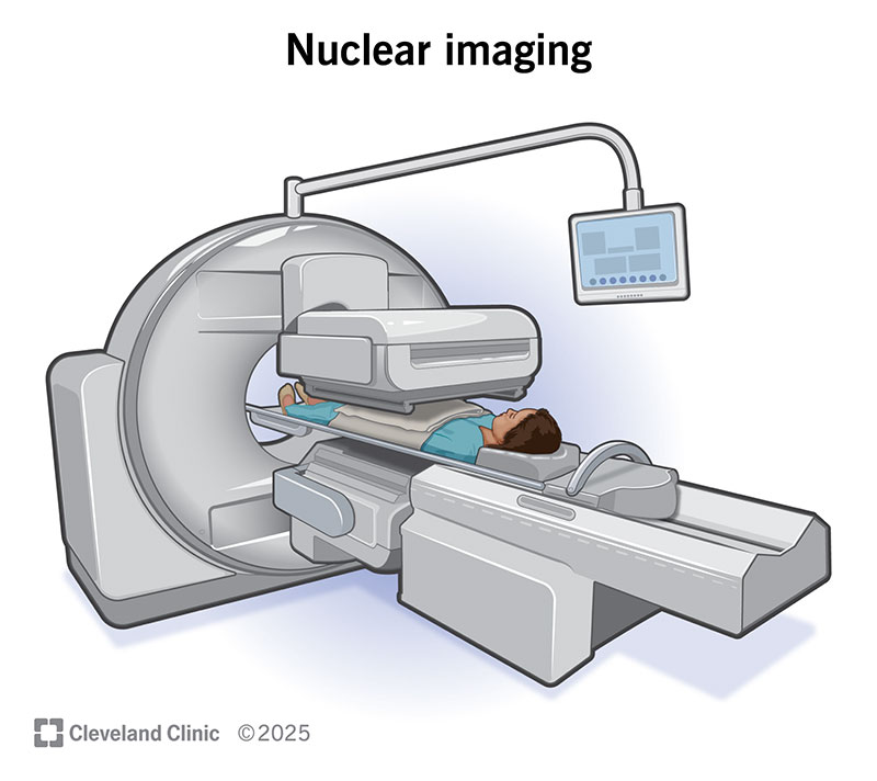
Image content: This image is available to view online.
View image online (https://my.clevelandclinic.org/-/scassets/images/org/health/articles/4902-nuclear-medicine-imaging-2.jpg)
Nuclear medicine imaging can see how well your internal organs and tissues are working.
It generally goes like this:
How you prepare depends on the kind of nuclear imaging test you’re having. Your healthcare provider will tell you what you need to do. Preparations could involve:
Advertisement
Always be sure to tell your healthcare provider about any allergies you have or medications you take. This can help them decide if the test is safe for you.
Here’s what you can expect after the tracer has time to circulate:
It depends on the type of test you’re having. In general, the scans might last about 30 to 60 minutes. This doesn’t include the time that it takes the tracer to travel. For scans involving bones, absorbing the tracer can take several hours.
It’s normal to feel concerned when you hear the word “radioactive.” But nuclear imaging is safe. It generally doesn’t cause side effects. The amount of radiation you receive is low. But it’s higher than tests like X-rays.
Some of the risks of nuclear medicine imaging are:
The benefits usually outweigh any risks of the test. Nuclear imaging can:
You can go back to your usual activities after the test. You typically don’t need to stay at the hospital. You can go home right after. You can eat and drink normally right away, too. Drinking water can help flush the tracer from your body more quickly. The tracer should be completely gone from your system within 48 hours.
It depends. A nuclear medicine doctor reviews the images from your scan. They create a report and send it to your healthcare provider (the one who ordered the test). Your healthcare provider usually discusses the results with you at a follow-up appointment. But they may give results over the phone. You can also read your results in your electronic medical record. This process can take a few days.
Be sure to ask your healthcare provider any questions you have about your results and what they mean for you.
Advertisement
Your healthcare provider will explain what they found on the test and what it means. If the test shows something irregular, they’ll discuss next steps. Sometimes, you need more tests to further investigate the problem. This could include:
If you have a nuclear imaging test, contact your provider if you experience any of the following after:
Having a nuclear imaging test may sound scary at first. But it’s one way your healthcare provider can see your organs and tissues and how they’re working. You’ll receive a safe, small amount of radioactive tracer. Then, a special camera will take pictures of your body. The tracer helps highlight problems and issues.
Your provider uses the pictures to diagnose things like infections, diseases and even cancer. It can also show how well certain treatments are working. Ask your provider any questions you have before the test. They can help you feel comfortable and confident about it.
Advertisement


Sign up for our Health Essentials emails for expert guidance on nutrition, fitness, sleep, skin care and more.
Learn more about the Health Library and our editorial process.
Cleveland Clinic’s health articles are based on evidence-backed information and review by medical professionals to ensure accuracy, reliability and up-to-date clinical standards.
Cleveland Clinic’s health articles are based on evidence-backed information and review by medical professionals to ensure accuracy, reliability and up-to-date clinical standards.
When you need a clear picture of what’s happening inside your body, the Cleveland Clinic imaging team is here for you.
