Atelectasis happens when the air sacs in your lungs can’t inflate properly, which means your blood, tissues and organs may not get oxygen. The most common cause is anesthesia during surgery. But pressure outside of your lung, a blockage or scarring may also cause it. It usually goes away after treating the underlying cause.
Advertisement
Cleveland Clinic is a non-profit academic medical center. Advertising on our site helps support our mission. We do not endorse non-Cleveland Clinic products or services. Policy
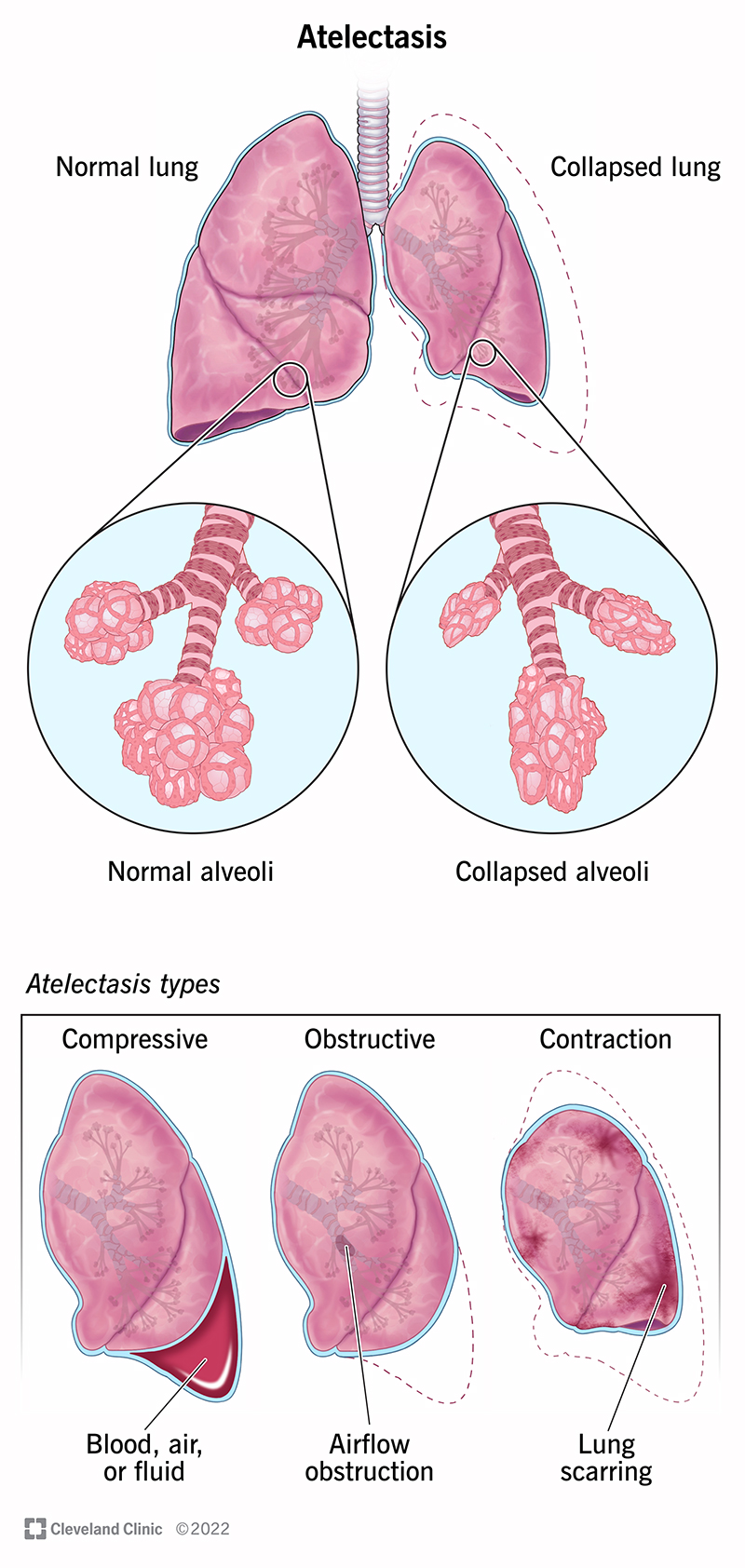
Image content: This image is available to view online.
View image online (https://my.clevelandclinic.org/-/scassets/images/org/health/articles/17699-atelectasis)
Atelectasis is when part or all of your lung deflates or collapses. Your lung is like a collection of balloons (air sacs, or alveoli) that fill with air. Atelectasis (pronounced “at-uh-LEK-tuh-sis”) occurs when some of these balloons lose air. It’s a common condition that may occur due to:
Advertisement
Cleveland Clinic is a non-profit academic medical center. Advertising on our site helps support our mission. We do not endorse non-Cleveland Clinic products or services. Policy
When you breathe in (inhale), your lungs fill with air. Air travels to the alveoli, where oxygen moves into your blood. Your blood delivers oxygen to organs and tissues throughout your body. But if your alveoli don’t get enough air, it can affect how well you breathe. This affects how much oxygen circulates through your body.
A collapsed lung sounds scary. And in some cases, it can be severe and even life-threatening. But atelectasis often goes away on its own. Depending on the cause, though, you may need treatment, like respiratory therapy, medications or a medical procedure.
Four of the most common types of atelectasis include:
Advertisement
Providers may also use different names to describe the location, appearance and severity of the alveoli collapse. These may include:
It usually doesn’t cause symptoms, although it may reduce your blood oxygen levels. Some causes of atelectasis can cause symptoms like:
Surgery is the most common cause of atelectasis. Healthcare providers use anesthesia to keep you asleep during surgery. Normally, you’ll take deep breaths or cough without thinking. This helps keep your alveoli open. But when you’re under anesthesia, you don’t breathe deeply enough to completely fill your lungs. You also don’t cough to clear your lungs of mucus. This can lead to blockages that prevent air from filling your alveoli.
Other atelectasis causes include:
Without treatment, atelectasis complications may include:
A healthcare provider will review your health history, perform a physical exam and order chest X-rays. They may recommend additional tests to help make an official diagnosis. These may include:
Most cases of mild atelectasis go away without treatment. But you’ll need regular appointments with your provider so they can monitor you closely.
Depending on the cause and how much your lung collapses, your provider may recommend:
Advertisement
Atelectasis after surgery usually goes away after 24 hours. Walking around, breathing exercises and using an incentive spirometer can help.
Other causes may take more time according to your treatment. Your healthcare provider will give you a better idea of what to expect.
Reach out if you have breathing problems, especially after surgery or if you have an underlying condition. Even what might seem like minor symptoms may be signs that your lungs aren’t working the way they should.
In most cases, with proper diagnosis and treatment, the outlook is good. It’s often reversible once healthcare providers determine the cause. Recovery is usually quick, and there aren’t any serious lasting effects.
If a long-lasting condition causes atelectasis, you may need further treatment. Your provider will tell you what to expect.
For most people who get atelectasis, it isn’t life-threatening. The condition usually goes away on its own or with proper diagnosis or treatment. But it’s still a good idea to talk to a provider any time you notice breathing changes, especially after surgery. They can answer any questions you have and recommend the best treatment.
Advertisement


Sign up for our Health Essentials emails for expert guidance on nutrition, fitness, sleep, skin care and more.
Learn more about the Health Library and our editorial process.
Cleveland Clinic’s health articles are based on evidence-backed information and review by medical professionals to ensure accuracy, reliability and up-to-date clinical standards.
Cleveland Clinic’s health articles are based on evidence-backed information and review by medical professionals to ensure accuracy, reliability and up-to-date clinical standards.
Breathing issues can affect your life in many ways. Cleveland Clinic’s respiratory therapy program treats your symptoms so you can feel better day to day.
