What is Ventricular Tachycardia?
The ventricles are the heart’s two lower chambers. They fill with blood from the top chambers of the heart (atria) and send it to the lungs and through the aorta to be circulated throughout the body. Tachycardia is a heart rate of greater than 100 beats per minute. A normal heart rate is 60 to 100 beats per minute at rest. Ventricular tachycardia is a regular, faster-than-normal heart rate that begins in the heart’s lower chambers. In most patients with ventricular tachycardia the rate is in the range of 170 beats per minute or more.
Causes
Your heart rate is controlled by electrical signals transmitted across the heart muscle. When something goes wrong and signals are sent too rapidly, tachycardia can occur.
Most often, ventricular tachycardia is caused by other heart problems such as coronary artery disease, high blood pressure, an enlarged heart (cardiomyopathy) or heart valve disease. It also can develop after a heart attack (myocardial infarction) or after heart surgery because of scar tissue that forms on the heart.
Ventricular tachycardia can develop in people who do not have heart disease, but this is less commonly the case. In these individuals, the condition can be caused by certain medications, an imbalance in electrolytes (the minerals that regulate heart rhythm), excessive caffeine or alcohol consumption, recreational drugs or exercise, or certain genetically transmitted conditions. In others, it occurs in the absence of heart disease or any other clearly identified causes.
Older people and those with a family history of heart rhythm disorders are more likely to develop ventricular tachycardia.
Symptoms
During an episode of ventricular tachycardia, the heart is beating so fast that the blood pressure drops so the heart cannot pump enough oxygen to every part of the body, and this is what causes symptoms. Although some people with ventricular tachycardia do not experience any symptoms, the most common symptoms are dizziness, lightheadedness, palpitations, shortness of breath or chest pain. When the heart rate is extremely high or the ventricular tachycardia persists for more than a few seconds, it can cause fainting, unconsciousness or cardiac arrest and death.
If you experience unexplained fainting, dizziness, lightheadedness, shortness of breath or palpitations, you should be evaluated for possible ventricular tachycardia. Chest pain, difficulty breathing and a rapid pulse are urgent symptoms of a potentially fatal ventricular tachycardia, and you must seek emergency care immediately to avoid the risk of cardiac arrest and death.
Heart Rhythms on EKG
The heart’s electrical system triggers the heartbeat. Each beat of the heart is represented on the electrocardiogram (EKG or ECG) by a wave arm.
Normal Heart Rhythm
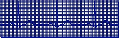
The normal heart rhythm (normal sinus rhythm) shows the electrical activity in the heart is following the normal pathway. The rhythm is regular and the node is normal (about 50 to 100 beats per minute).
Tachycardia
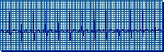
Tachycardia: fast heart rhythm (> 100 beats per minute)
Diagnosis
The most common first step in diagnosing ventricular tachycardia is an electrocardiogram (ECG/ EKG). An EKG is a device used to record on graph paper the electrical activity of the heart. The picture is drawn by a computer from information supplied by electrodes (small sticky patches) that are placed on your chest and arms to record the heart’s rhythm. The doctor will perform an EKG in the office, but you also may need to wear a portable EKG unit (Holter monitor) at home for 24 to 48 hours so the doctor can better evaluate your heart’s rhythm.
Another tool to help diagnose ventricular tachycardia is electrophysiology testing. Your doctor may refer you to a specialist to have this procedure done. During the test, catheters with electrodes are inserted through the veins at the groin and placed directly in the heart to study the electrical activity of different regions of the heart. The doctor can then create a map of the heart’s electrical impulses and determine precisely where the abnormalities originate.
Treatments
The goals of treating ventricular tachycardia are to manage any underlying disease, which may improve or eliminate the abnormal heart rhythm, and prevent future episodes.
In an emergency, the immediate goal is to slow the heart rate to normal. This may involve CPR, electrical defibrillation or giving IV medications.
Most patients with ventricular tachycardia have two treatment options — radiofrequency catheter ablation (RCA) and an implantable cardioverter defibrillator (ICD). Both treatment options are associated with good outcomes when they are performed at a medical center where the doctors are highly experienced in treating patients with ventricular tachycardia.
Radiofrequency Catheter Ablation
Radiofrequency catheter ablation (RCA) is a procedure performed by a cardiac electrophysiologist, which is a cardiologist who focuses on the treatment of heart rhythm disorders. In the first part of the procedure, the physician uses electrophysiology techniques to pinpoint the location in the heart where the abnormal rhythm originates.
In the second step, the physician uses a catheter with a special tip on it that emits a high frequency form of electrical current. The energy is directed at the area in the ventricle where the abnormal current originates to destroy a tiny amount of tissue. This is called an ablation procedure.
Ablation of ventricular tachycardia has a long history of safety and success. For some patients, ablation completely cures the abnormal rhythm, and they require no further treatment. For other patients, ablation enhances the effectiveness of an ICD.
Ablation of Ventricular Tachycardia
Cleveland Clinic is a national referral center for patients with ventricular arrhythmias, performing more than 500 ablation procedures for this condition from 2003 through 2009.
In 2009, Cleveland Clinic physicians performed 95 ablations for ventricular tachycardia and eliminated all tachycardias in 82 percent of those patients. In another 6 percent, the procedure successfully eliminated at least one tachycardia in patients with multiple tachycardias.
Implantable Cardioverter Defibrillator
An implantable cardioverter defibrillator (ICD) is a device that monitors and controls the heart’s rhythm to prevent ventricular tachycardia. It is implanted under the skin.
An ICD consists of a pulse generator, which is about the size of a pager, and one or more lead wires that connect the pulse generator with the heart. The leads are inserted through the veins and positioned in the heart. The leads transmit the heart’s electrical activity to a computer microchip in the pulse generator. If the computer detects an abnormal heartbeat, the pulse generator sends an electrical signal across the lead(s) to the heart to restore normal heart rhythm.
Clinical studies show that ICDs are effective for nearly all patients in stopping life-threatening ventricular tachycardias.
Medications to slow down the heart rate are another treatment option for patients with ventricular tachycardia. These drugs can be effective but are associated with some serious, potentially fatal side effects, and their use is declining.
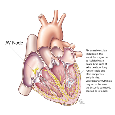
The Heart's Electrical System

The atria (the heart’s upper chambers) and ventricles (the heart’s lower chambers) work together, alternately contracting and relaxing to pump blood through the heart. The electrical system of the heart is the power source that makes this possible. Here’s what happens during a normal heartbeat:
- Sinoatrial (SA) Node
With each heartbeat, the electrical impulse begins at the SA node, located in the right atrium. The SA node produces the electrical impulses that set the rate and rhythm of the heartbeat. The electrical activity spreads through the walls of the atria and causes them to contract. - Atrioventricular (AV) Node
The AV node is located between the atria and ventricles and acts like a gate that slows the electrical signal before it enters the ventricles. This delay gives the atria time to contract before the ventricles do. - HIS-Purkinje Network
This pathway of fibers sends the impulse into the muscular walls of the ventricles and causes them to contract. This contraction forces blood out of the heart to the lungs and body.
The SA node fires another impulse and the cyle begins again.
Follow-Up Care
If you are under a doctor’s care for ventricular tachycardia, it is important that you keep your appointments for regular follow-up visits. Your doctor will want to monitor your condition, make sure the treatment is still effective and discuss any symptoms or other changes in your condition that may occur.
It is important for you to continue to follow a heart healthy diet and exercise plan as recommended by your healthcare team.
Appointments
By Phone
To make an appointment, please call 800.223.2273 or 216.444.6697.
International patients, please call Global Patient Services at 001.216.444.8184.
To talk with a nurse about ventricular tachycardia and available treatment options, please contact a heart, vascular & thoracic resource center nurse toll-free at 866.289.6911.
Online
MyConsult® Online Medical Second Opinion
MyConsult Online Second Opinion program connects patients to the expertise of top Cleveland Clinic specialists without the time and expense of travel. Through our secure web platform, patients can submit their detailed health information, medical records and diagnostic test results. The most appropriate Cleveland Clinic expert is assigned to the consultation and will render a detailed second opinion. The report includes commentary about the diagnosis and treatment options or alternatives and recommendations regarding future therapeutic considerations. Patients are also able to send additional questions to the physician who provided the report. Online medical second opinions are available for more than 1,200 medical diagnoses.
Chat with a Heart Nurse
For over 15 years Heart, Vascular & Thoracic resource nurses have been offering assistance to people who have questions or concerns about heart, vascular and thoracic conditions. Nurses are available to help with questions on symptoms, diagnoses, treatment options, Cleveland Clinic services and doctors, etc. The nurses are available weekdays from 8:30 a.m. – 4 p.m., Eastern Time, through phone, secure live chat or email. If you need help, you may contact a nurse.
Why Choose Us?
About the Miller Family Heart, Vascular & Thoracic Institute
The Sydell and Arnold Miller Family Heart, Vascular & Thoracic Institute at Cleveland Clinic is one of the largest cardiovascular specialty groups in the world, providing patients with expert medical management and a full range of therapies.
Our areas of expertise combine research, education and clinical practice to provide innovative and scientifically based treatments for cardiovascular disease. The commitment of our physicians and scientists to the prevention and cure of cardiovascular disease has led to innovative care, better outcomes and improved quality of life for patients with cardiovascular disease.
Resources
Learn More
- Learn more about ventricular tachycardia by visiting Cleveland Clinic’s Heart, Vascular & Thoracic Institute website
- Watch videos on heart rhythm disorders and treatment presented by Cleveland Clinic specialists
- Read posts about ventricular tachycardia on our daily blog, Health Essentials from Cleveland Clinic
- Sign up for our e-newsletter, and get tips on maintaining a heart healthy lifestyle, recipes and essential health news
