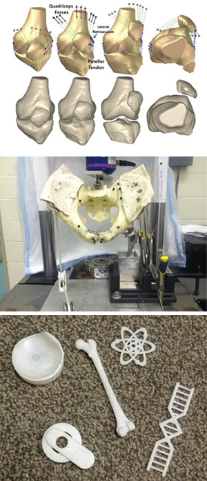
The Cleveland Clinic Akron General Biomechanics Laboratory combines medical imaging with biomechanical analysis to evaluate how anatomy, injury and surgical treatment influence joint function. The Biomechanics Laboratory focuses on collaborative studies including physicians, scientists and engineers throughout Cleveland Clinic. The Biomechanics Laboratory is a component of the Cleveland Clinic Program of Advanced Musculoskeletal Imaging and Musculoskeletal Research Center.
The Biomechanics Laboratory has a specific focus on injuries that hinder function of the patellofemoral joint, such as patellar dislocations and patellofemoral pain. The patient-focused research approach combines quantitative MRI, anatomical analysis, biomechanical modeling, and outcomes analysis to evaluate mechanisms of injury, treatment strategies and long-term influence on function. Evaluating mechanisms of injury improves understanding of the functional characteristics that cause patellofemoral disorders.
Treatment strategies are studied to evaluate how surgical and conservative treatment options influence joint mechanics. Evaluation of long-term function primarily focuses on progressive cartilage degradation to osteoarthritis triggered by a patellofemoral disorder. The overall goals of the program are to reduce the risk of injury and optimize patient-specific treatment strategies to return patients to an active lifestyle while also preserving patellofemoral cartilage.
The key resources within the laboratory include two material testing machines, video based and electromagnetic 3D tracking systems, a thin film pressure measurement system, device fabrication facilities including a 3D printer, and facilities for anatomical dissection.
Several workstations are available with software for computer aided design, construction of graphical models from MRI and CT, 3D manipulation of the models, technical computing, multibody dynamics analysis, and statistical analysis of data.
Imaging data is acquired for multiple sites within Cleveland Clinic and external collaborators. Funding for projects within the laboratory is obtained from a wide variety of grants and contracts from federal agencies and foundations.

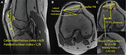
Measurements used to characterize knee anatomy and patellofemoral alignment.

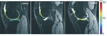
T1ρ relaxation times mapped to patellofemoral and tibiofemoral cartilage after a patellar dislocation.

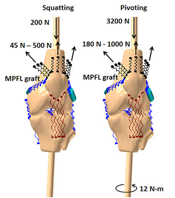
Computational models for simulation of knee function.