Echocardiography – What you need to know
Subscribe: Apple Podcasts | Buzzsprout | Spotify
Echocardiography – What you need to know
Podcast Transcript
Announcer: Welcome to Love Your Heart, brought to you by Cleveland Clinic's Sydell and Arnold Miller Family Heart & Vascular Institute. These podcasts will help you learn more about your heart, thoracic, and vascular systems, ways to stay healthy and information about diseases and treatment options. Enjoy.
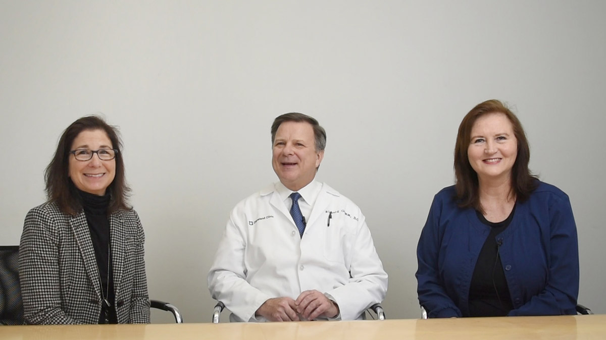
Moderator: I'm here today with Dr. Rick Grimm, who is the medical director of our echo lab, and Helga Lombardo, who's the manager of our echo lab, to talk about echocardiogram. So Dr. Grimm, can you talk about the different types of echo, and then how do doctors choose the type of echocardiogram for a patient?
Dr. Rick Grimm: Sure. So, an echocardiogram is just an ultrasound procedure specifically geared towards evaluating the heart, hence the term echocardiogram. Can also be called cardiac ultrasound. But it's a non-invasive test. And it's very routinely performed and, in fact, I would consider it to be the most important diagnostic test for cardiologists, for physicians in general, in terms of wanting to evaluate all aspects of the heart relative to heart function, valvular disease, hemodynamics and concern for artery disease. So it's a very, very important test to us.
Patients will see different environments in which these are done and our environment, our laboratory, is actually associated, in fact, proximity wise in the ground floor of our hospital, and we actually perform these tests in our laboratory, on both inpatient and outpatients alike on any given day.
It's relatively simple and is absolutely painless for the patients. The patients come into our laboratory, they do need to be put into a gown in order that we are able to access the chest wall, and a small probe is actually inserted on the chest wall. And from the surface of the chest, we are able to acquire the ultrasound images. And the test generally takes between 30 and 60 minutes, depending on the complexity of the case and the underlying diagnosis of the patient. But it, again, is a procedure in which the patient simply needs to lie on a bed and the images are acquired, and after about 30 to 60 minutes, most of the information is usually obtained on any given case.
Moderator: So sometimes patients have to get a more extensive echo, there might be ones with exercise or they may get ones where there's a probe down their throat...
Dr. Rick Grimm: Right.
Moderator: ... or different things, how do you choose which one?
Dr. Rick Grimm: Right, right. Well, first of all, regarding the one that we actually insert like an endoscope, and just like a gastroenterology examination of the stomach, the probe, the actual imaging capability is on the end of this probe, and we can insert this into the food pipe. It sits in the esophagus, which is right next to the heart, and we're able, as a result, to get very, very high definition, high fidelity images from the esophagus because we're so close to the heart, and very exquisite images of the heart. As opposed to the chest wall where the ultrasound beam needs to penetrate through the bone, and skin and soft tissue, again, when it's sitting in the esophagus right next to the heart, the images are just much more high fidelity and high resolution images.
And so, in some cases, in the overwhelming majority of the cases, we can acquire adequate images from the chest wall, but in some cases, and particularly for structures of the heart that are posterior, or in the back aspect of the heart, this transesophageal imaging is optimal. And often, for example, patients that have any pathology of the mitral valve, which is a more posterior structure, or the left atrium, which is a more posterior structure in the heart, that is best imaged with a transesophageal echocardiogram.
Moderator: Okay. And what should patients know as far as coming for an echocardiogram?
Dr. Helga Lombardo: For a standard transthoracic echocardiogram, there really isn't much to the patient. It's a painless test. They'll be lying on a table, asked maybe to have their positions changed from one lateral decubitis to on their back. They may hear sounds, which is the doppler coming out of the machine, but for the most part it's a painless test.
For a stress echo, if they're coming for a stress echo, to wear comfortable shoes, because they'll be running on a treadmill. And for a transesophageal echo, basically again, they're transferred into a gown, but they do need to bring a driver because they are lightly sedated, and they need to make sure that they have someone who can safely take them home.
Moderator: A lot of times patients try to read their tests on their own. And they'll call because they have questions, what would you like to say to patients about the test results?
Dr. Rick Grimm: Yeah, right. Well, I would caution the attempt to interpret without some guidance. A lot of the terminology can be very involved and complex of course, and potentially misleading unfortunately. And some factors that might appear somewhat unusual or out of the range of normal, etc., may or may not be in fact, depending on what exactly is being evaluated. In many cases, it may not even be pertinent to the given patient's condition.
So I would interpret those with a grain of salt, when you're looking at that. And ask questions of your physician, of course, and directly address medical personnel regarding those findings and any questions you may have, but definitely.
Moderator: Well, thank you both for answering our questions...
Dr. Helga Lombardo: Thank you.
Moderator: ... and I know this will be very helpful to our patients.
Dr. Rick Grimm: Wonderful, thank you.
Dr. Helga Lombardo: Thanks.
Moderator: Mm-hmm (affirmative)
Announcer: Thank you for listening. We hope you enjoyed the podcast. We welcome your comments and feedback. Please contact us at heart@ccf.org. Like what you heard? Please subscribe and share the link on iTunes.
Love Your Heart
A Cleveland Clinic podcast to help you learn more about heart and vascular disease and conditions affecting your chest. We explore prevention, diagnostic tests, medical and surgical treatments, new innovations and more.