A Look at Echocardiography

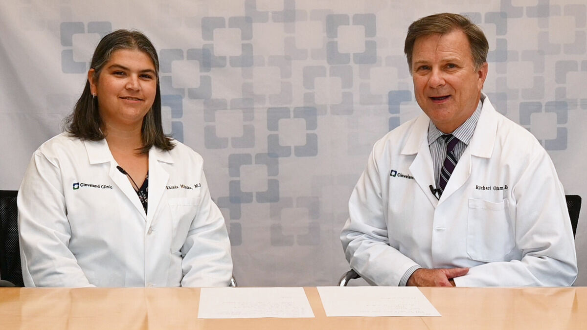
Echocardiography can be a useful diagnostic tool in caring for cardiac patients. Dr. Richard Grimm and Dr. Rhonda Miyasaka speak about various types of tests using echocardiography. Learn indications, benefits, and challenges of these imaging tests.
Subscribe: Apple Podcasts | Buzzsprout | Spotify
A Look at Echocardiography
Podcast Transcript
Announcer:
Welcome to Cleveland Clinic Cardiac Consult brought to you by the Sydell and Arnold Miller Family Heart, Vascular and Thoracic Institute at Cleveland Clinic.
Richard Grimm, DO:
Good afternoon. My name is Rick Grimm, and I'm here with one of my partners here at Cleveland Clinic, Dr. Rhonda Miyasaka. Rhonda is one of my colleagues in the cardiac, Cardiovascular Imaging Section here at Cleveland Clinic. Rhonda heads up our structural intervention team and is a critical member of our group. I'm the Director of the Echocardiography Laboratory, as well as our medical information officer here at Cleveland Clinic. And we're here to talk to you about echocardiography and the utility in your patient population and hopefully to answer some questions and concerns that you may have relative to obtaining an echocardiographic test in 2021 here. As I'm sure many of you are familiar and aware, echocardiography is a critically important, non-invasive technology for all of us in the diagnostic medicine, diagnostic cardiology realm. It's critically important to not only diagnose the presence of cardiac dysfunction, but significant valve disease and certainly congenital anomalies as well.
Richard Grimm, DO:
The field of echocardiography just continues to expand and just become greater and greater in terms of its utility, its capabilities, the innovation that's been afforded to the area, as well as the image quality in our capabilities expanding to other realms as stress echocardiography, transesophageal echocardiography, in the interventional realm, as well as in the operating room to really help us guide procedures as well. It's often asked of us when we field questions from our colleagues as to what might be the best test to offer. For example, a transthoracic echocardiogram versus a transesophageal echocardiogram. And as one of our transesophageal echo experts, I'll ask Rhonda to address that issue and perhaps try and help make it more clear in terms of ordering these tests.
Rhonda Miyasaka, MD:
Sure. So we have, as you all know, several different options when we're doing echocardiography, and that includes a transthoracic echocardiogram or a transesophageal echocardiogram. And one of the really common questions that we get in the Echo Lab is, "Which one should I order? Should I start with a transthoracic echo and then get a transesophageal echo?" Or sometimes, "Should I just skip right to the transesophageal echo and not worry about the transthoracic echo?" And so something that I like to say is that these two imaging tests are very much complementary. And so the transesophageal echo is very good at looking at valves in detail, looking at valvular function, looking at valvular structure, and we get really, really fine images. If we need to look for endocarditis, if we need to look for blood clots in the heart or the appendage, transesophageal echo, as you all know, is excellent.
Rhonda Miyasaka, MD:
Transthoracic echo also has things that it's better at compared to transesophageal echo. And I think that's something that we sometimes forget. We think that because a TEE is a more invasive procedure and we get closer to the heart, we think that that's just going to give us everything. In fact, a transthoracic echo also can give us a wealth of information, particularly if we're trying to understand LV size, function, wall motion, or if we're looking at images that would be in the far field, close to the apex on transesophageal echo. When we think about where our transthoracic echo probe is, it's right close to the apex. And so that's why, depending on what part of the heart we're looking at, sometimes transthoracic echo actually does a better job. So typically, when I'm thinking about a patient and what imaging test to start with, I almost always want to start with a transthoracic echo so that we can get a sense of you what our image quality is.
Rhonda Miyasaka, MD:
We can get a sense of what we're able to see and what we're not able to see. And then when we do our transesophageal echo, if we need to, we're able to go with very focused information gathering so that we know ahead of time some of the things that we might be looking for. So I think that's kind of one of the main things that I always like to point out is that the two tests are very much complimentary, and don't forget about transthoracic echo. We actually have amazing power these days. We can do 3D transthoracic echo. We can do 3D of the valves. We can do all of our measurements. So that's something that we're very happy with.
Richard Grimm, DO:
The way I like to think of it, sometimes in terms of the utility and especially when it comes to measuring or sampling blood flow and gradients and so forth, from the chest wall, you have an unlimited number of frames of reference to actually interrogate flows. And obviously, in order to get the most accurate data, we have to be well-aligned with that flow. So we can do that relatively easily. It's a challenge technically in any case, but we can do it much, much better from the chest wall. And again, we have an infinite number of locations in which to interrogate from. When we're in the esophagus with a transesophageal probe, of course, we're pretty limited. We have an infinite number of alignments that we can perform. But the position and the angulation is limited, obviously, because we can only go so far. One of the other tests that's extremely useful for us in the Echo Lab is the stress test.
Richard Grimm, DO:
And typically, stress testing is used for the evaluation of coronary artery disease and ischemia. But in fact, in our laboratory and we do a lot of nuclear studies as well as a lot of stress echocardiograms in our department, we find it extremely useful, especially in patients with valvular heart disease. So that we can evaluate the response of the heart muscle in a given state in terms of severity of valve disease and the response of that heart muscle to stress. Obviously, if all things are relatively good, that heart muscle is going to respond favorably. If it doesn't, we can see evidence of that. We can measure sample gradients that peak stress and actually make an assessment as to the potential impact of that valvular heart disease on that heart muscle. So that's an extremely valuable test, not only in terms of acquiring that data, but also the data relative to functional capacity, heart rate, blood pressure, et cetera, as well. So again, another extremely useful test. And maybe Rhonda, you could speak to the stress that we utilize treadmill and dobutamine.
Rhonda Miyasaka, MD:
Sure. A common question that we get is, "Well, I think my patient needs a stress test, but what kind should I order? Should I order a treadmill echo or should I order a dobutamine echo?" And so I can talk about kind of the pros and cons of both approaches. In general, if the patient is able to exercise and if we're trying to understand symptoms, for example, that come on with exercise, it's always better to try to do a treadmill because that's physiologic. If the patient mentions they're having shortness of breath or chest pain when they're walking or exercising, then we can try to simulate those conditions with exercise. And we can see if they develop chest pain. We can see if there are abnormalities we notice on echo. And so it often gives us a lot of additional information. In addition to just looking at the echo images, we get a lot of functional information from the test as well.
Rhonda Miyasaka, MD:
If a patient is not able to walk, if a patient just broke their leg and we're not going to exercise them on a treadmill, then in that situation, a dobutamine stress test is a way that we can still do the stress test. We can simulate the heart under stress and look for signs of ischemia, but we can't get quite as much information potentially from the symptoms that the patient's experiencing because it's not really a physiologic state. We also will sometimes use dobutamine to evaluate a valve disease. That's something that, from time to time, we'll do with aortic stenosis, for example, if we have to tease out some of the subtleties of whether the aortic stenosis is severe or not. But those are kind of the finer points of when we might use dobutamine. But in general, if your patient's able to exercise, we prefer an exercise test.
Richard Grimm, DO:
And one last mention just regarding the lab and what's done on a routine basis increasingly in the world of echocardiography and learned this many years ago, that the importance of quantification is key. And certainly, other techniques, other modalities, whether it's CT and cardiac MRI, obviously give us exquisite images and exquisite accuracy in the echo world as well. We've really upped our game relative to the quantification of both function as well as valve severity. And the importance of that, of course, everybody can understand. But that's a significant emphasis currently, and it's something that we expect to improve significantly in the very near future as well.
Rhonda Miyasaka, MD:
Yeah, I think that's kind of a nice summary of what to expect when we order an echo. And hopefully that was helpful for everyone.
Richard Grimm, DO:
Wonderful. Thank you for joining us.
Announcer:
Thank you for listening. We hope you enjoyed the podcast. We welcome your comments and feedback. Please contact us at heart@ccf.org. Like what you heard? Subscribe wherever you get your podcasts or listen at clevelandclinic.org/cardiacconsultpodcast.

Cardiac Consult
A Cleveland Clinic podcast exploring heart, vascular and thoracic topics of interest to healthcare providers: medical and surgical treatments, diagnostic testing, medical conditions, and research, technology and practice issues.


