Overview
Caring for your retina
When you open your eyes in the morning, chances are you don’t think about your retina. You might not even know what your retina is, what it does or where it’s located in your eye. But the retina is one of the most important parts of your eye — allowing you to see the world around you. A thin, light-sensitive lining in the back of your eye, the retina contains millions of special nerve cells that react to light. These cells (photoreceptors) send electrical impulses to your optic nerve, which your brain converts into the images you see.
Most people don't worry about the health of their retina until something goes wrong. But retinal diseases are the leading causes of blindness in adults in the United States.
Seeking treatment as soon as possible is often critical when it comes to many retinal diseases. Your eyes are your windows to the world — expert care can help prevent vision loss and keep your eyes as healthy as possible.
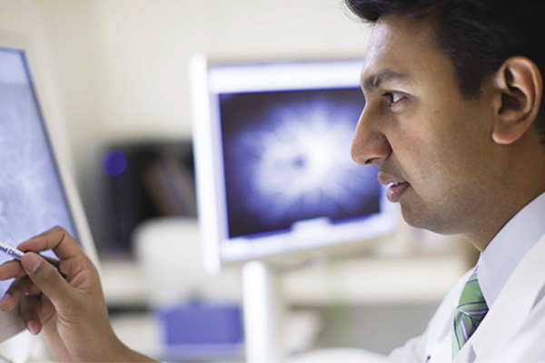
Why choose Cleveland Clinic for retinal disease care?
Cleveland Clinic is recognized in the U.S. and throughout the world for its expertise and care. Our retina specialists are internationally recognized for their expertise and world-class, customized care. Our team uses the most innovative approaches, state-of-the-art imaging technologies and equipment to diagnose and treat the entire spectrum of retinal disorders—from the routine to the complex. At Cole Eye Institute, you’ll have access to our entire team of specialists and subspecialists to ensure you receive the best treatment, specially tailored just for you.
Many procedures developed at Cleveland Clinic have been adopted by ophthalmologists worldwide. Our robust and ongoing research continues to expand our understanding of eye disease, giving us some of the best clinical outcomes in the field. This research also provides patients who qualify with access to the latest clinical trials and new treatments that might not be available elsewhere.
What We Treat
- Age-related macular degeneration (AMD).
- Central serous chorioretinopathy.
- Cystoid macular edema.
- Diabetic retinopathy.
- Eye cancer (retinoblastoma).
- Inherited retinal dystrophies.
- Macular hole.
- Macular pucker.
- Posterior vitreous detachment.
- Retinal artery occlusion.
- Retinal detachment / retinal tear.
- Retinal inflammatory disease.
- Retinal vein occlusion.
- Retinopathy of prematurity.
Diagnostic Testing
At Cleveland Clinic, we take the time to accurately evaluate and diagnose your condition. Our healthcare providers work with you to determine what's going on within your eye. Some of the tests we use can include:
- Optimal coherence tomography (OCT): This noninvasive, painless test takes a cross-section picture of your retina. It's one of the most common diagnostic imaging tests.
- Spectral domain optical coherence tomography (SDOCT): The newest, highest resolution OCT imaging technology, SDOCT displays greater detail of your retinal layers.
- Interoperative OCT: Using OCT technology in the operating room offers the eye surgeon high-resolution views of your retinal tissues as they are manipulated during surgery.
- Fluorescein angiogram (FA): This test involves taking a rapid series of photos (not X-rays) of your eye, while a small amount of yellow dye is injected into your arm. The dye causes the blood vessels to stand out and will show if there are any changes to your retina.
- Indocyanine green (ICG) angiogram: Like the fluorescein angiogram, this test also involves injecting a dye (green this time) into your arm, while a rapid series of photos (not X-rays) are taken of the choroid (deeper layer of your eye just behind your retina). The dye causes the choroid blood vessels to stand out and will show if there are any changes to your choroid.
- Ultra-widefield fundus ttechnology: This painless, noninvasive imaging technique offers a more detailed and enhanced view of your entire retinal periphery in just one image.
- Bright scan ultrasound (B Scan): Painless and noninvasive, the B Scan uses sound waves to bypass an obstruction, such as a cataract, retinal tear or vitreous hemorrhage (blood in the eye). that hinders a clear view of the back of your eye.
- Ophthalmic electrophysiology: This series of tests goes beyond standard eye exams to measure how different parts of your eye respond to light. These tests can help diagnose genetic disorders of the retina and are also used to measure optic nerve disease, toxicity to the eyes from medication or metallic foreign bodies and active inflammation.
Our Doctors
Looking for a retina specialist?
Find a ProviderRetinal Appointments & Locations
To make an appointment with one of our ophthalmologists, please call 216.444.2020.
