The Sydell and Arnold Miller Family Heart, Vascular & Thoracic Institute is one of the largest cardiovascular practices in the United States. With so many patients, our caregivers have collectively seen it all and experienced it all.
What does that mean to you? It means that as a patient of Cleveland Clinic, you’ll have access to the broadest possible range of solutions from skilled, experienced doctors, nurses and technicians. Options you may not have in your hometown – or anywhere else in America.
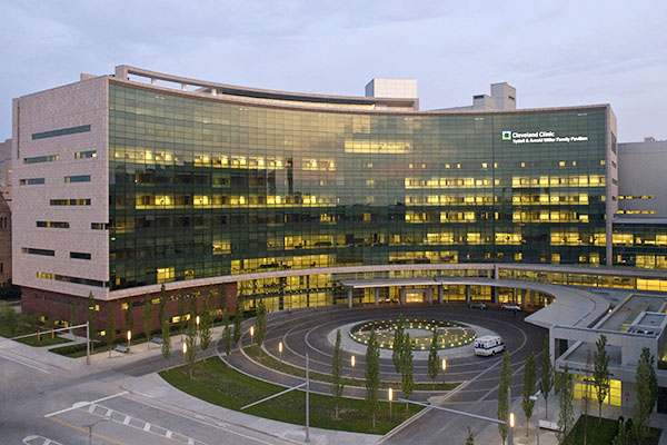
About the Miller Family Heart, Vascular & Thoracic Institute
Treatment Outcomes
Download details about our treatment outcomes for surgical and other types of care.
Traveling for Heart Surgery
Patients travel from all over the world to have heart surgery at our nationally and internationally ranked program. Discover why.
Patient Stories
Real stories from patients who have received care from The Sydell and Arnold Miller Family Heart, Vascular & Thoracic Institute.
Recognition & Awards
Cleveland Clinic is nationally and internationally renowned for its cardiovascular care.
Giving
Help us continue to provide world-class heart and vascular care to patients from all over the world.
Community Outreach
Programs aimed at educating and improving heart health in our community.
Patient & Family Health & Education Center
Computers, educational handouts, and classes available to patients and family members.
