What is the Retina?
Your retina is the light-sensitive lining in the back of your eye. It contains millions of special nerve cells that react to light. These photoreceptors send electrical impulses to your optic nerve, which your brain converts into the images you see.
Most people never give their eyes — let alone their retinas — a second thought until something goes wrong. Yet, retinal diseases are the leading causes of blindness in adults in the United States. Common retinal diseases include age-related macular degeneration (AMD), diabetic retinopathy and retinal detachment. More uncommon conditions include retinal inflammatory disease and retinitis pigmentosa.
Seeking treatment as soon as possible is often critical when it comes to many retinal diseases. Don’t delay. In many cases, early diagnosis and treatment can help stop vision loss.
Macular Degeneration

In the United States, age-related macular degeneration (AMD) is the No. 1 cause of legal blindness in adults. One of several types of macular degeneration, AMD occurs when the small central portion of the retina (the macula) is diseased and the eye’s ability to distinguish fine details is affected.
Most patients have the “dry” form, in which yellow deposits (drusen) are present in the macula. Drusen may be asymptomatic or accumulation may cause dimming or distortion. In advanced stages of dry AMD, the disease can cause blind spots and central vision loss.
About 10 percent of AMD patients develop the “wet” form, in which abnormal blood vessels grow from the choroid and under the retina. The vessels leak blood and fluid into the retina, distorting vision (making straight lines look wavy), and creating blind spots and central vision loss.
How is AMD treated?
There is no cure for AMD, but early detection and treatment can delay or reduce its severity. Cole Eye Institute’s retina staff works with patients and their families to determine the best treatment option:
- Vitamins — A combination of vitamins C, E, lutein, zeaxanthin, zinc and copper have been shown to decrease the risk of vision loss in patients with intermediate to advanced dry AMD or wet AMD in one eye.
- Anti-vascular endothelial growth factors drugs — Currently used as firstline therapy for wet AMD, the results of clinical trials of local injections of ranibizumab (Lucentis®), afibercept (Eylea®) and bevacizumab (Avastin®) for active wet AMD showed visual stability in about 95 percent of patients and visual gain in about 35 percent of patients.
- Photodynamic laser therapy — This involves the injection of a light-sensitive drug into the bloodstream. After being absorbed by the abnormal blood vessels, the drug is activated with a cold laser, which destroys the unwanted blood vessels in select wet AMD cases.
- Laser therapy — High-energy light is occasionally used for select subtypes of wet AMD to destroy active growing abnormal blood vessels.
Diabetic Retinopathy
An eye disease that affects people with diabetes, diabetic retinopathy occurs as a result of high blood glucose, or sugar, that diabetics often have over a prolonged period of time. Too much blood glucose can damage the blood vessels in the back of the eye, preventing the retina from receiving the proper amount of nutrients it needs to maintain vision. In most cases, patients do not notice a significant change in their vision until late in the disease.
Diabetic retinopathy occurs when diabetes damages the tiny blood vessels in the retina. In the early stages of the disease (non-proliferative retinopathy), these blood vessels may leak fluid, resulting in edema. In the more advanced stage (proliferative retinopathy), fragile new blood vessels grow in the retina and in the vitreous humor (a clear substance that fills the eye). If left untreated, these blood vessels may bleed and cloud vision, or may scar and detach the retina.
How is diabetic retinopathy treated?
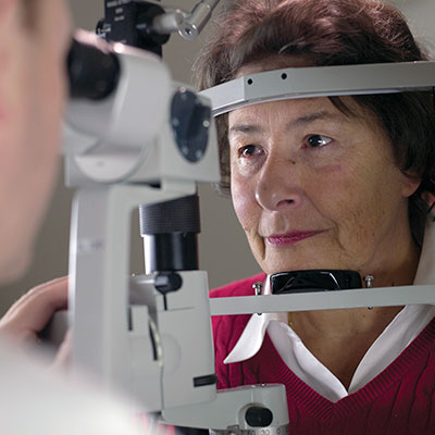
Regular monitoring of the retina with dilated fundus exams and leading-edge imaging technologies (such as optical coherence tomography) is important in diabetic retinopathy. If detected at earlier stages, more than 90 percent of patients can be saved from blindness. Other treatments include:
- Medications — Now becoming the first-line treatment for many types of diabetic eye disease, these medications include anti-vascular endothelial growth factor inhibitors (such as ranibizumab, afibercept and bevacizumab), steroids (such as dexamethasone intravitreal implant, or Ozurdex®), fluocinolone acetonide intravitreal implant (Iluvien®) and triamcinolone acetonide injectable suspension (Triescence®). Studies show that stability and improvement in visual acuity appears to be more significant with these therapies.
- Laser surgery — In many cases, laser surgery reduces the risk of future vision loss. Laser photocoagulation can be performed to seal or destroy growing or leaking blood vessels in the retina.
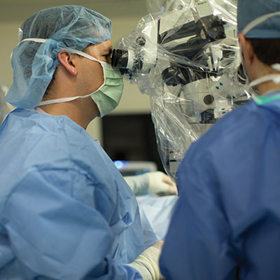
- Vitrectomy — In some people with diabetic retinopathy, the blood that leaks from blood vessels in the retina also may leak into the vitreous humour, clouding vision. These vessels also may cause scar tissue and subsequent retinal detachment. Vitrectomy can be used to remove the blood or repair a retinal detachment. At Cole Eye Institute, our specialists typically perform sutureless small-gauge surgery that increases patient comfort and speeds recovery.
Be sure to schedule follow-ups with your primary care physician and/ or endocrinologist to optimize systemic control of blood sugar, blood pressure and blood lipids.
Retinal Detachment

Retinal detachment is a serious condition that occurs when the retina pulls away from its supporting tissues. Since the retina cannot work properly under these conditions, permanent vision loss can occur if a detachment is not repaired quickly.
Nearsightedness, previous trauma, family history of retinal detachment and previous eye surgery increase risk for retinal detachment, but it may also be spontaneous.
How is retinal detachment treated?
There are a number of approaches to treating a detached retina, including:
- Laser (thermal) or freezing (cryopexy) — If diagnosed early enough, both of these approaches can repair a retinal tear before it causes retinal detachment.
- Pneumatic retinopexy — Repairs select retinal detachments. Cryo treatment is first used to seal the tear. A small gas bubble is injected into the vitreous, where it rises and presses against the retina, closing the tear.
- Scleral buckle — This procedure involves placing a silicone band (buckle) around the eye to hold the retina in place. This band is not visible and remains permanently attached. Cryo treatment closes the tear. A gas bubble is often used to help reattach the retina. This procedure has a 95 percent effective rate.
- Vitrectomy — Used for large tears, the vitreous is removed from the eye and replaced with a saline solution. A gas or silicone oil bubble and laser are used at the time of vitrectomy to facilitate repair. Patients will need to maintain a face-down position for a period of time to keep the bubble in place. Success rates are similar to the scleral buckle. In some cases, both scleral buckle and vitrectomy are indicated to repair a retinal detachment.
Macular Hole
A macular hole occurs when the nerve cells of the macula become separated from each other and pull away from the back surface of the eye. Sometimes macular holes are the result of an injury or a medical condition that affects the eye. In most people, it seems to be a side effect of the changes that normally occur in the eye as we age.
How is macular hole treated?
Our experts work with patients to determine whether surgery — which is usually the recommended treatment — or watchful waiting is preferable. In select cases, using an injection of ocriplasmin (Jetrea®) is recommended.
Surgery involves vitrectomy (removal of the gel-like vitreous fluid from the eye), as well as removal of any pieces of tissue near the macula. The fluid in the eye is replaced with a sterile gas, which keeps pressure on the macular hole until it heals. Patients will need to maintain a face-down position for a period of time to keep the gas bubble in place. Medical therapy using a novel enzyme (such as ocriplasmin) also is available for treating select macular hole.
Success rates of macular hole anatomic closure reached 99 percent at Cole Eye Institute over the past few years. We use the latest small gauge surgical techniques to improve patient comfort and decrease surgical time.
Macular Pucker
The macula normally lies flat against the inside back surface of the eye. Sometimes cells can grow on the inside of the retina, contracting and pulling on the macula. Occasionally, an injury or medical condition creates strands of scar tissue inside the eye. These are called epiretinal membranes, and they can pull on the macula. When this pulling makes the macula wrinkle, it is also called macular pucker. In some eyes, this will have little effect on vision, but in others it can be significant, leading to distorted vision.
Sometimes macular puckers are the result of an injury or a medical condition, such as diabetes, that affects the eye. Epiretinal membranes can sometimes form after eye surgery. The cause of most cases of macular pucker is not known.
How is a macular pucker treated?
Cole Eye Institute retina experts work with patients to determine whether macular puckers should be closely monitored or treated with surgery.
Surgical treatment includes a vitrectomy to remove the gel-like vitreous fluid from the eye. The surgeon also will peel the membranes away from the macular surface. This should allow the macula to lie flat against the back of the eye and improve visual symptoms.
Retinal Inflammatory Disease
Retinal inflammatory disease includes a wide variety of conditions that cause problems with vision. These conditions may be limited to the eye (such as inflammation caused by a virus or fungus), or may be part of a disease that affects multiple organ systems (such as autoimmune disorders). It may be rapidly progressive, making it difficult to treat. Retinal uveitis also may be caused by viruses including those related to shingles or herpes, bacteria (such as tuberculosis or syphilis), or parasites (such as toxoplasmosis).
How is retinal inflammatory disease treated?
Cole Eye Institute retina doctors work closely with patients to diagnose retinal inflammatory diseases. Treatments vary depending on the cause of the inflammation and may include medications or surgery. Treatments are targeted at the particular diagnosis, such as antibiotics for a bacterial infection or steroids for a primary inflammatory disorder.
We have extensive experience in managing such retinal inflammatory diseases to minimize their impact on quality of life and to manage relapses. Retina surgeons at Cole Eye Institute also have extensive experience in the use of long-acting implants (steroidal) in the treatment of these conditions, such as the Retisert® and Ozurdex® implants.
Pediatric Retinal Diseases
Retinal diseases affect children of all ages. In premature babies, a number of factors, including exposure to oxygen and low birth weight lead to a process by which abnormal blood vessels can lead to retinal detachment and blindness if not detected and treated properly. This disease is called retinopathy of prematurity (ROP).
How is ROP treated?
Fortunately, premature babies at risk for ROP in newborn nurseries are examined for its presence. Cole Eye Institute pediatric retina specialists are active in screening babies for this disorder.
Laser treatment is effective in preventing retinal detachment and vision loss. Babies who do not respond may need additional vitreoretinal surgery. In addition to diagnosing and treating patients with ROP, Cole Eye Institute physicians and researchers are investigating the underlying cases of this disease in the laboratory to improve outcomes.
How are systemic diseases in children treated?
Systemic diseases in children can often have retinal complications, including diabetes, sickle cell anemia, inflammatory disorders, neurodegenerative diseases, and a variety of inherited metabolic syndromes.
Cole Eye Institute pediatric specialists are experienced in detecting retinal complications of these diseases, collaborating with pediatricians and other physicians, and providing therapy as needed.
How are retinal injuries treated?
Children are commonly involved in injuries that could result in vitreous hemorrhage and/or retinal detachment. Members of Cole Eye Institute’s vitreoretinal team are able to operate on such children and reattach the retina, improving chances for visual recovery.
Can retinal tumors affect children?
Retinal tumors, although rare, also can affect children. Retinoblastoma is the most common type in pediatric patients. Cole Eye Institute’s ocular oncology staff has extensive experience in the diagnosis and management of children with retinoblastoma and collaborates, when needed, with experts from Cleveland Clinic Taussig Cancer Institute.
Appointments
Ready to Make an Appointment with a Specialist?
If you would like to set up a consultation with a Cleveland Clinic Cole Eye Institute retina specialist, please call 216.444.2020 or 800.223.2273. In many instances, same-day appointments for new patient and follow-up visits are available.
Virtual Second Opinion
If you cannot travel to Cleveland Clinic, help is available. You can connect with Cleveland Clinic specialists from any location in the world via a phone, tablet, or computer, eliminating the burden of travel time and other obstacles. If you’re facing a significant medical condition or treatment such as surgery, this program provides virtual access to a Cleveland Clinic physician who will review the diagnosis and treatment plan. Following a comprehensive evaluation of medical records and labs, you’ll receive an educational second opinion from an expert in their medical condition covering diagnosis, treatment options or alternatives as well as recommendations regarding future therapeutic considerations. You’ll also have the unique opportunity to speak with the physician expert directly to address questions or concerns.
Why Choose Us?
Cleveland Clinic Cole Eye Institute offers expert retinal care
At Cleveland Clinic Cole Eye Institute, our retina staff has the expertise to accurately diagnose and offer world-class treatment for retinal diseases, macular diseases including age-related macular degeneration, diabetic retinopathy and retinal detachment. We also treat more uncommon conditions, such as retinal inflammatory disease and retinitis pigmentosa.
Cole Eye Institute is among the world’s most advanced eye institutes. Our fully integrated model helps us provide patients with quick and easy access to specialty and subspecialty care for a wide spectrum of eye conditions — from the routine to the complex.
All care at Cole Eye Institute is provided in the most patient-friendly and effective way. Each year, our internationally recognized staff carries out more than 140,000 patient visits and performs more than 8,000 surgeries — volumes among the highest in the nation.
The Cole Eye Institute has a reputation for innovation and superior outcomes, and its research team is dedicated to understanding eye disease in hopes of finding tomorrow’s cures.
By choosing Cole Eye Institute, you can take comfort in knowing you have quick and easy access to our entire team of specialists and subspecialists should you require additional vision care.
As a patient, you will not only benefit from our clinical experience; you also have the advantage of an active research team, which bridges the gap between laboratory research and patient care, and offers access to the latest clinical trials, should you qualify. These research studies not only provide treatments not otherwise available, but they also help us expand our overall understanding of eye disease.
Please use this guide as a resource as you examine your treatment options. Remember, it is your right as a patient to ask questions and to seek a second opinion.
What Cleveland Clinic offers
Thorough evaluation and accurate diagnosis is critical to receiving the most appropriate treatment. At Cole Eye Institute, we offer the most state-of-the-art imaging technology available, including:
- Spectral Domain Optical Coherence Tomography (OCT) — This latest generation of imaging technology (15 times more sensitive than conventional ultrasound) provides high-resolution information regarding retinal and ophthalmic tissue anatomy, facilitating diagnosis and guiding management.
- Fluorescein Angiography (FA) — A technique for examining the retina’s circulation uses a dye tracing method.
- Indocyanine Green (ICG) Angiography — A special dye test to evaluate the circulation of the choroid, the layer just behind the retina.
- Ultra Widefield Fundus Photography and Angiography — A special imaging technique that allows for visualization of the far retinal periphery, assisting in diagnosis and management.
- Intraoperative OCT — Integrating OCT technology in the operating room that allows for high-resolution anatomic visualization of tissues during surgical procedures and image-guided surgical interventions.
- OCT Angiography — It is now possible to image retinal blood vessel patterns with OCT technology.
Center for Genetic Eye Diseases
Genetically determined retinal dystrophies and degenerations are one of the leading causes of congenital blindness in developed countries. They also can appear later in childhood and the teenage years. Some examples of these diseases include Leber congenital amaurosis, Stargardt disease and retinitis pigmentosa.
Cole Eye Institute provides a specialized retinal dystrophy clinic with advanced diagnostics, genetic counseling and genetic testing. Some of the patients examined at Cole Eye Institute have gone on to receive gene therapy for their disease.
Ophthalmic Imaging Center
Leading-edge imaging technologies have transformed the clinical and surgical care for vitreoretinal diseases. Cole Eye Institute’s Ophthalmic Imaging Center houses leading research programs in novel imaging technologies, such as intraoperative optical coherence tomography. The Ophthalmic Imaging Center includes vitreoretinal specialists, engineers and researchers focusing on translating these technologies to patient care.